


Characteristics of thoracic osteochondrosis
This type of disease is quite rare compared to the cervical and lumbar spine.
- It is the longest (consisting of 12 vertebrae);
- There is a slight natural curvature in this area - physiological kyphosis - that relieves some of the load that occurs when walking upright;
- The thoracic vertebrae are connected to the ribs and sternum, perform the functions of the physiological framework and bear the main load;
- In cross section, the spinal canal in the thoracic region has the smallest dimensions;
- The thoracic vertebrae are thinner and smaller in size, but have longer spinous processes.
Stages of thoracic osteochondrosis
- The first stage is characterized by disc dehydration, which causes the disc to lose elasticity and stiffness but retains the ability to bear normal loads. The discs begin to flatten, lose height, and form bulges. The pain at this stage is mild.
- In the second stage, cracks form in the annulus fibrosus and instability of the entire segment is recorded. The pain becomes more intense when bending and doing other movements.
- The characteristic sign of stage three is that the annulus fibrosus ruptures and begins to form a herniated disc.
- During the transition to stage four, due to the lack of resistance in the intervertebral discs, the vertebrae begin to move closer together, causing spondyloarthrosis (disorder of the intervertebral joints) and spondylolisthesis (twisting or shifting of the vertebrae). Mobilization of compensatory forces to reduce load causes the vertebrae to grow, increase in area, and flatten. The affected portion of the annulus fibrosus begins to be replaced by bone tissue, which significantly limits the movement capacity of this sector.
Degree of thoracic osteochondrosis
Typical signs and symptoms
- Intercostal Neuralgia - Pain that is often concentrated in one area and then spreads rapidly throughout the chest, forcing the patient to maintain a certain position and significantly complicating breathing.
- The pain becomes more severe with turning, neck movements, bending, raising arms, breathing movements (inhale-exhale).
- Severe spasms in the muscles of the mid and upper back. Muscle fibers in the abdominal muscles, lower back, and shoulder girdle may also contract, which is reflexive in nature (occurring as a response to sharp pain syndrome).
- Intercostal neuralgia typically causes pain, stiffness, and discomfort in the chest and back during activity. The pain can be severe, last for several weeks without further spread, and then begin to subside.
- All symptoms become more pronounced at night. In the morning they soften or subside considerably, are aggravated by hypothermia, movement (especially vibrations and sudden movements), and may manifest themselves in some stiffness.
Atypical signs and symptoms
- Mimics the pain characteristics of heart disease (heart attack, angina). This pain can be quite persistent (unlike heartache), and traditional medications used to dilate coronary blood vessels do not eliminate the pain. The electrocardiogram also showed no changes.
- In the acute phase of thoracic osteochondrosis, there is often prolonged (up to several weeks) sternal soreness reminiscent of breast disease. They can be ruled out by examination by a breast doctor.
- Abdominal (ilium area) pain similar to colitis or gastritis. When located in the right rib cage, cholecystitis, pancreatitis, or hepatitis are often misdiagnosed. These symptoms often accompany disruption of the digestive system due to compromised innervation. In this case, it is necessary to identify thoracic osteochondrosis as the primary disease causing such manifestations.
- If the lower chest area is damaged, the pain is localized in the abdominal cavity, simulating intestinal pathology but independent of the quality of food intake and diet. Increased pain severity is primarily due to physical activity.
- Reproductive or urinary disorders can also occur due to distortions in the innervation of organs.
- Damage to the upper thoracic segment will cause symptoms such as pain in the esophagus and pharynx, and foreign body sensation in the pharyngeal cavity or behind the sternum.
Back pain and back pain
- back; back
- Back pain.
- The duration can reach 14-20 days;
- Exacerbation of the syndrome is observed when bending to the sides, forward, or taking deep breaths;
- When upper back pain occurs, movement in the cervicothoracic area is limited; when lower back pain occurs, movement in the lumbar and thoracic area is limited;
- The pain worsens at night and may disappear completely when walking;
- Deep breathing and staying in one position for long periods of time can cause the pain to worsen.
diagnosis
- Radiography. With its help you can detect:
- Changes in the anatomy of the damaged segment;
- Thickening of intervertebral discs;
- Vertebral body deformation and displacement;
- Differences in disc height.
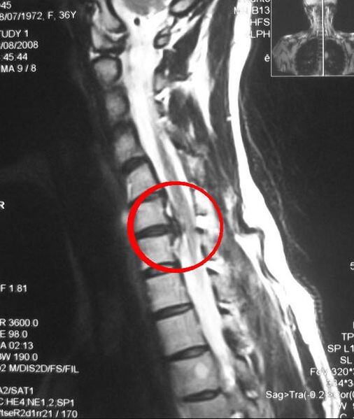
- Computed tomography (CT) and magnetic resonance imaging (MRI) are more accurate methods because they provide layer-by-layer images of the affected area.
- Electromyography is performed to differentiate neurological symptoms due to nerve root compression in thoracic osteochondrosis. You should get checked if:
- Impaired motor coordination;
- Headache;
- Dizziness;
- Pressure fluctuations.
- Laboratory tests - determine calcium levels and ESR (erythrocyte sedimentation rate) in the blood.